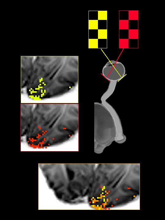
Functional neuroimaging, psychophysical, and electrophysiological investigations were performed in a patient with non-decussating retinal-fugal fibre syndrome (Apkarian et al., 1994a, 1995), an inborn achiasmatic state in which the retinal projections of each eye map entirely to the ipsilateral primary visual cortex. Functional magnetic resonance imaging (fMRI) studies showed that for monocularly-presented simple visual stimuli, only the ipsilateral striate cortex was activated. Within each hemisphere's striate cortex, the representation of the two hemifields overlapped extensively. Despite this gross miswiring, visual functions that require precise geometrical information (such as vernier acuity) were normal, and there was no evidence for the confounding of visual information between the overlapping ipsilateral and contralateral representations. Contrast sensitivity and velocity judgements were abnormal, but their dependence on the orientation and velocity of the targets suggests that this deficit was due to ocular instabilities, rather than the miswiring per se. There were no asymmetries in performance observed in visual search, visual naming, or illusory contour perception. fMRI analysis of the latter two tasks under monocular viewing conditions indicated extensive bilateral activation of striate and prestriate areas. Thus, the remarkably normal visual behavior achieved by this patient is a result of both the plasticity of visual pathways, and efficient transfer of information between the hemispheres.
