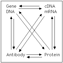
Go to: dictionary | proteins | cDNAs | DNA &
genes | bioinformatics | logic
& exptl design
(home page) |
GPIN
home Page
 Scope and
objective
Scope and
objective
Scope and objective. Biological scientists have used antibodies for many years to study proteins; but, as is the case with protein purification and recombinant DNA technology, the ways antibodies are produced and used has led to an increasingly powerful technology. The purpose of this section is to discuss the way these antibodies are produced and how they can be used. We will skirt the basic biology of the immune system as much as possible, but we must note that the discovery of how organisms have developed methods to generate millions of potential antibodies reacting with millions of potential antigens is an intellectual triumph that would bring delight to any biological scientist. The combination of using multiple alleles which undergo specific recombination events and are further modified by somatic cell mutation is a fascinating story and the way the body controls this process will certainly be an object of study for decades to come, but these topics are appropriate to a course devoted to immunology.
Antibodies. Antibodies* actually include several classes of molecules some of which like the IgA are designed for secretion in the bodily fluids while others, like the IgM are designed to be expressed on the cell surface. The antibody that is most useful in biological studies is the IgG class, a protein molecule that is made and secreted and can recognize specific antigens. The IgG is composed of two subunits including two "heavy" chains and two "light" chains. These are assembled in a symmetrical structure and each IgG has two identical antigen recognition domains. The antigen recognition domain is a combination of amino acids from both the heavy and light chains. The molecule itself is roughly shaped like a "Y" and the regions of the extreme tips of the "Y" are the antigen recognition domains. Limited protease digestion of the antibody produces two 'Fab' fragments, each of which has a single antigen binding site. Thus, an intact IgG can cross-link two antigens, which may have important biological implications, while a Fab fragment could not do this. For example, cross linking cell surface antigens with an IgG can cause a change in their distribution on the cell surface or stimulate internalization. The stem of the "Y" is not involved in recognition of antigens and is fairly constant among the various classes of antibodies. This region is called the Fc region and is important because it itself can be used as an antigen that is recognized by other antibodies and because it binds with high affinity to the A protein which is a protein that can be isolated from Staph Aureus. The antigen-recognizing regions of the molecule are concentrated in the Fab regions. It is possible to treat antibodies with proteases to cleave the antibody near the hinge region and produce the monovalent Fab fragments which must be used if there is a need to avoid dimer formation.
Antigens. The proteins that can be recognized (bound) by the antibody are called antigens*. These antigens are most commonly polypeptides or carbohydrates, but they can also be lipids, nucleic acids, or even small molecules like neurotransmitters. A particular antibody molecule can only interact with a small region of an antigen and in the case of a polypeptide this is generally about 5-12 amino acids. This region can be continuous or it can be distributed in different regions of a primary structure that are brought together because of the secondary or tertiary structure of the antigen. The region of an antigen that is recognized by an antibody is called an epitope*. It is possible, although unlikely, that the same, or closely related epitopes, could be shared by the different antigens. In some cases, it is possible to make an antibody that is directed against the antigen binding site of another antibody (i.e., the antigen binding site is the epitope). This type of antibody is called an anti-idiotype antibody.* In this case, the antigen binding site of the anti-idiotype antibody can be similar in structure to the original antigen (they both recognise the same antibody.)
Adding an epitope: If a cDNA for a protein is available, it is possible to add a new coding sequence before expression, producing a fusion protein. Since only a few amino acids are required to form an epitope*, only a few amino acids need to be added to a protein to produce a strong antigen binding site. This forms an epitope-tag, and common versions of the epitope tag are FLAG-tag*, His-tag, and GST-tags.
Thinking about antibody antigen interactions. It is important to be constantly aware of the way antibodies recognize antigens and the nature of the antibodies that are used. It is possible to use serum as a source of antibody but in this case the antibodies that are present are derived from many independent clones each of which produces a separate antibody. (Serum is the fluid that is produced when blood clots.) In this case, multiple antibodies may recognize multiple epitopes on a single protein which can be useful experimentally.
In other cases antibodies can be produced by a single clonal cell line (called monoclonal antibodies) and in this case every antibody molecule is identical to every other one and all recognize the same epitope. This is an important distinction to remember since using both sera and monoclonal antibodies have advantages and disadvantages. It is clearly easier to think about how a monoclonal antibody can be recognized, but it must be remembered that since any single antibody molecule recognizes only a single epitope, it is always possible that this epitope may be shared among very different proteins. So this must be considered in experimental design. This problem is partially avoided by the use of antisera which are by their nature polyclonal. An antisera that has been produced with a high titer to a particular antigen will have many antibodies recognizing many epitopes on that antigen. Because of this it is less likely that a cross-reacting epitope could be a problem. On the other hand, antisera include not only antibodies directed against a particular antigen of interest, but they may also include many other types of antibodies present in the serum (yes, they are always there). These antibodies may be present due to the immunological history of the animal, or even the presence of an autoimmune reaction. Experimentally it is essential that the experimentalist considers the possibility that reacitivities present in antiserum are due not only to reaction to an antigen of interest but due to some other antigen. Thus, controls, including the use of the preimmune serum are always essential. Frequently, using both an antiserum and the monoclonal antibody for a specific experiment can help make a convincing case.
Making polyclonal antibodies (antiserum). Historically, serum was the first source of antibodies. In some cases, a serum may have a high concentration of an antigen of interest without any experimental manipulation. Good examples of this are the existence of human autoimmune antisera that are produced by patients with Lupus or multiple sclerosis or paraneoplastic disorders. When such a serum is available it provides an experimental tool that can be used to study the underlying immune response and to isolate and study the antigen or the cDNA for the antigen that is recognized by the antibody. A number of antisera have serendipitously recognized particular antigens of interest. For example, there is one antiserum that specifically stains neuronal precursors in Drosophila embryo. More commonly, it is necessary to experimentally produce a high titer antisera.
an antiserum is made by injecting an antigen into an animal, most commonly a rabbit or a chicken (sometimes to bypass the problems of tolerance) but also hamsters, rats, goats, and even cows. The quality of the antisera produced will be determined in part by the quality of the antigen selected. An antigen can be either a purified protein, or a relatively complicated mixture of several macromolecules. Antisera have been produced, for example, against preparations of secretory granules (which contain multiple proteins and other antigenic macromolecules). Such an antisera is obviously complicated, but it has proved useful to some investigators.
If the sequence of a protein is available, it is also possible to generate a synthetic peptide which can be covalently attached to a "carrier" protein and used as an antigen to make an anti-peptide antibody.* Typically, an antigen is mixed with an adjuvent mixture and injected into an animal. Adjuvents are frequently chosen in an attempt to increase the strength of the immune response, but there is much debate about which adjuvents are effective or even if they are effective at all. After an initial injection an animal undergoes a primary immune response which produces a relatively low titer antiserum and includes antigens of multiple classes including IgM and IgG. After a waiting period, the animal is injected again (boosted) to produce a secondary response. This procedure can be repeated multiple times, but each time the injection procedure is repeated, the repertoire of antibodies produced by animal is somewhat different. Thus, every antiserum has a slightly different specificity and some scientists are apt to guard their pool of "good" antisera for fear that the fraction they have may be all that will ever be produced. The serum is produced by withdrawing blood from the animal. The blood is allowed to clot removing red blood cells, white blood cells, and some serum proteins. The remaining fractions is a heterogeneous mixture that includes many proteins including the antibodies of interest. In some cases the serum is further purified before use. It is possible to absorb an antisera with material in an attempt to increase the specificity of the antiserum. For example, an antiserum directed against brain synaptic vesicles might be absorbed with a microsome fraction from liver in an attempt to increase specificity. There are also straight forward procedures to purify the IgG fraction from serum so that potentially interfering substances in the serum can be removed. Finally, the antibodies that specifically recognize an antigen of interest can frequently be purified using an affinity column that contains the substance (peptide, carbohydrate, protein, etc.) that was used as an antigen initially. The quality of an antiserum of course must be judged on the basis of specific assays and these will be discussed below.
Making monoclonal antibodies. An important advance in the use of antibodies was the recognition that it was possible to produce a monoclonal population of antibodies. This is a procedure that is in many ways analogous to isolating a cDNA from a cDNA library. In this case, an animal produces a library of B cells that are secreting different antibodies. The task of the experimentalist is to select a single B cell that is producing the antibody of interest and to expand that population so that a large amount of secreted antibody can be produced. This experimental task was successfully accomplished by Kohler and Milstein (Nature 256:495 (1975)) in the late seventies and rapidly became part of the standard repertoire of biological scientist. B cells of an immunized animal can be isolated in high concentration from the spleen of that animal but antibody producing cells by their nature are short-lived and cannot be easily cloned and expanded. This problem was solved by a procedure that involves fusion with an immortalized B-cell line. This fusion is done by standard cell fusion approaches (e.g., mixing the cells and concentrating them in the presence of polyethylene glycol). The line that is chosen to fuse with the B cells has several properties. First, it is an immortalized line which means in many cases a fusion between that cell and a B cell will produce an immortal cell type. Second, the line is usually chosen so that it does not produce an antibody itself. Third, it is chosen with a selectable marker so that after the fusion is completed the parental cells can be removed.
Most commonly this selectable marker is the enzyme HGPRTase*. It is possible to select for cells deficient in this enzyme with the agent 6-TG, which is toxified by HGPRTase. It is also possible to select for the presence of HGPRTase using the HAT* selection. In this protocol, Aminopterin (HAT) is used to block de novo synthesis of purines. Hypoxanthine (HAT) is providers of purine precursor. Only if HGPRTase is present, will hypoxanthine be incorporated into purines. Since Aminopterin also prevents synthesis of thymidine (HAT), exogenous thymidine must also be present. Thus, B-cells are isolated, fused with an immortalized line, and the hybrid cells are selected. B-cells die because they are not immortal and the HGPRTase-deficient lines die because of HAT selection. A diagram illustrating this proceedure. is here.
The next, and most experimentally challenging, part of the procedure is to design a method of selecting (i.e., cloning) an individual cell producing an antibody of interest. Most commonly, this is done by a limiting dilution approach. The culture is divided into many individual smaller cultures and the supernatant of each culture is then sampled and assayed for the antibody of interest. This assay can be anything from a radio immunoassay to an assay dependent on the ability to identify a protein after a western analysis or to recognize a particular cell using immunohistochemistry. The key component of the assay is that it be specific (that it recognize the antigen or antigens of interest) and that it be rapid enough that it can be experimentally convenient. Once such an assay is established, individual wells that contain the antibody of interest can be identified and the cells present in that well can be isolated and re-plated at even lower dilutions. The assay can then be repeated and the isolation and dilution can be repeated until a clonal population is isolated. Of course, it is essential that a clonal population be isolated or the reagent will be heterogeneous. If two cells are present, these cells may be producing different antibodies and one may outgrow the other. In the latter case, if the cell of interest grows more slowly, it will eventually be lost in culture, so using a rapid assay is important.
The main advantages of isolating monoclonal antibodies are that (a) they are a homogeneous population that (b) they can be produced in essentially unlimited quantities. This means that the reagents can be carefully characterized and shared among multiple investigators and used for an extended period of time without change in quality. Monoclonal antibodies thus can serve as a common standard for scientists in widely different locations and can be incorporated into commercial applications that can be produced on an industrial scale. The main weakness of monoclonals is that they recognize only a single epitope, so there is always the possibility that an epitope that is shared by two antigens can confuse the experimentalist. An antigen directed against a carbohydrate can be shared by many proteins and even glycolipids.
Uses of antibodies. Antibodies have a wide variety of uses.
1. They can be used to immuno-localize a particular antigen in a tissue (immunohistochemistry*). Tissue can be fixed and incubated with the antibodies of interest. These antibodies can then be localized using a 'secondary' antibody coupled to a gold particle or another enzyme that gives a chemical reaction like horse radish peroxidase or beta galactosidase. A secondary antibody is frequently made by generating an immune response to the Fc region of the primary antibody in another species. Thus, if the primary antibody is a mouse IgG, then the secondary could be a rabbit anti-mouse antibody which has been linked to beta galactosidase. Alternatively the antibody can be purified and then conjugated to another molecule to produce a fluorescent antibody.
2. Antibodies can be used to quantitate the presence of an antigen using either a radioimmunoassay* (RIA) or an enzyme-linked immunoabsorbent assay (ELISA)*. There are many variants of these approaches, but those are based on a similar idea. For example, if an antigen can be bound to a solid support or surface, it can be detected by reacting it with a specific antibody and the antibody can be quantitated by reacting it with either a secondary antibody or by incorporating a label directly into the primary antibody. Alternatively, an antibody can be bound to a solid surface and the antigen added. A second antibody that recognizes a distinct epitope on the antigen can then be added and detected. This is frequently called a 'sandwich assay' and can frequently be used to avoid problems of high background or non-specific reactions. It was only an assay of this type, for example that was sensitive and reproducible enough to measure low concentrations of nerve growth factor present in the body.
3. Antibodies can have effects in vivo. These effects can be used in an experimental sense to determine the importance of a particular molecule in vivo. For example, antibodies to nerve growth factor demonstrated that nerve growth factors had to be present for the sympathetic and sensory nervous system to develop. Antibodies to particular hormones or cell-surface receptors can also have a therapeutic use. For example, antibodies to EGF receptor have effects on the development of some tumor types.
4. Since antibodies have a high affinity for a particular epitope, they can be used as affinity reagents in protein purification.
5. Because particular antibodies bind to proteins with high affinity they can also be used as a classic criteria for the importance of a particular enzyme or other macromolecule in a particular reaction. If an antibody can interfere with a reaction in a solution, that suggests that it may be binding specifically to a protein involved in that reaction. Of course, it must be remembered that some antibodies may be directed against a site on a protein that is not important for enzymatic activity and so may not have the classic "blocking" activity.
If an antibody can react with a specific protein, that protein can subsequently be precipitated from solution, frequently with the help of a secondary antibody that will cross-link the antibody-antigen complexes made. Alternatively, the antigen-antibody complex can be removed by reacting the solution with either protein A or an anti-Fc antibody which has been attached to beads so that can be easily removed form the solution. These approaches provide a powerful criterion for identifying and studying the function of proteins.
6. Antibodies can also be used in conjunction with gel-shift experiments to identify specific DNA-binding proteins. As discussed in the section on transcription analysis, DNA-binding proteins can be frequently assayed by their ability to bind with high affinity to a particular oligonucleotide. The mobility of an oligonucleotide associated with the protein is far different than the mobility of a free oligonucleotide giving a signal that is frequently called a gel shift. If an antibody to a protein present in such a complex is available, the addition of the antibody to the binding assay can have either of two effects. If the antibody binds to a region of a protein not involved in DNA binding it can produce a complex that has even a slower mobility leading to another shift in mobility (a super-shift). Alternatively, if the antibody binds to a region of the protein involved in recognizing the DNA then it can disrupt the binding and eliminate the shift. In either case, this can serve as a criterion to identify DNA-binding protein.
7. It is also possible to use an antibody to detect a protein after fractionation by SDS-PAGE. This approach, which is called 'western blotting*' because it is analogous with northern blotting and Southern blotting, involves transferred a fractionationed protein to a nitrocellulose sheet. These proteins can then be exposed to a particular antibody and the antibody can recognize the protein bound to the blot. This allows the identification of a particular protein. This approach is particularly useful if the mobility of the protein changes during an experiment. For example, incorporation of a phosphate, or a carbohydrate or cleavage of the protein results in a change in mobility and this can be followed in straight forward manner by western analysis. With appropriate controls, this approach can be use to measure the abundance of a protein in response to experimental manipulations.
8. The combination of SDS gels and immunoprecipitations* can also be extremely effective. If a particular protein can be immunoprecipitated in a solution, both supernatant and precipitated fractions can be separated on an SDS gel and studied.
9. Antibodies, including antisera, can be used to screen expression libraries to isolate candidate genes that express a particular epitope.
10. If a fluorescent antibody is bound to a cells surface it can be used as a marker to quantitate the fraction of cells expressing that marker using flow cytometry*. If different antibodies / fluorescent dye combinations are used, the fraction of cells expressing several antigens can be determined.
11. Anti-idiotype antibodies, which are antibodies against the binding domain of another antibody, can sometimes be used to mimic the structure of the original antigen.
Antibody Specificity. perhaps most importantly, it is important to consider the question of antibody specificity and reactivity. The quality of experimental work using an antibody cannot be any greater than the quality of the antibody that is used. Doing immunolocalization or immunoprecipitation with a poorly characterized antibody is more likely to add confusion than enlightenment to a field, but a well characterized antibody is invaluable. Once an antibody (serum or monoclonal) is produced, it may be characterized by a number of approaches. Does it precipitate an antigen of the correct size? Does it react with a single peptide by western analysis? Is the protein it recognizes present in the 'right' tissues (remembering that antigens are sometimes found in unexpected places)? If it is an anti-peptide antisera, is its reactivity competed by the peptide used to make it? (This is a good reason to make anti-peptide antiseras.) Is it localized where it might be expected by immunohistochemistry (tissue distribution or subcellular localization)? Does the antigen appear when the gene is expressed (either during development or with an expression vector)? Is the protein missing (or changed in its properties) in an animal with a mutation? All these are useful criteria, and it is incumbent on the experimenter to develop sufficient criteria that the experiments using an antibody are convincing.
Suitability of antibodies for different purposes. It should be appreciated that not all antibodies will work equally well in all assays. An antibody for example may work beautifully in a western style analysis but fail in immunohistochemistry. Thinking about the nature of antibodies in the epitopes they recognize, may explain this initially puzzling phenomenon. If an antibody is directed against an epitope that is buried in the interior of the protein, it may not be accessible in immunohistochemistry. Likewise, if a reagent used to fix a tissue modifies a residue that is a key residue in a particular epitope an antibody may not recognize a protein in a fixed tissue. Thus, immunohistochemical localization may depend not only on the quality of the antibody but also on the type and extent of fixation. If an epitope is on the surface of the protein and is made up by amino acids that are widely separated in the primary structure of the protein, then the denaturation of the protein on an SDS gel may irreversibly separate and disrupt this epitope. If one is interested in immunoprecipitating a native protein, it is presumably essential to have an antibody that recognizes a surface epitope on that protein with reasonably high affinity. If the affinity of the antibody is too low then the complex may dissociate before the particular insoluble fractions are separated. Despite these considerations, the use of antibodies is an important approach to studying cells and their proteins.